Torn Retinaculum : Achilles Tendon Rupture Treatment Carlsbad Ca Achilles Tendon Injury Oceanside Ca : This condition may be associated with tears of the peroneus brevis and or peroneus longus.
Torn Retinaculum : Achilles Tendon Rupture Treatment Carlsbad Ca Achilles Tendon Injury Oceanside Ca : This condition may be associated with tears of the peroneus brevis and or peroneus longus.. Stretching this ligament keeps the patella in place and the ligament healthy.stretching a lateral retinaculum of the knee. The medial retinaculum is located within the knee joint; This occurs because of the chronic pull of the knee cap to the outside by the thigh muscles, creating a strain on the medical or inside tissues (the retinaculum). The main symptoms of inferior extensor retinaculum pain are: In a lot of ways, our bodies are strung together like a puppet or marionette, the strings being our tendons, ligaments, and other connective fibers and tissues.
Most of the fibers of the medial patellar retinaculum originate in the medial femoral region from the vastus medialis muscle, just superior to the patella. The flexor retinaculum of the foot can be strained or injured due to a variety of reasons. An injury, rupture, or tear to any one of these strings can slow down or even incapacitate the body. Tear of the superior peroneal retinaculum at its attachment to the distal fibula Again a previously torn retinaculum will be thickened and ill defined compared to a normal retinaculum which is usually thin and well defined.
Peroneal Tendon Dislocation Dysfunction Vascular Health Clinics from www.vascularhealthclinics.org If this is your first visit, be sure to check out the faq & read the forum rules. The medial retinaculum plays a minor role — along with the vastus medialis oblique and the medial patellofemoral ligament — in providing medial stability in. The lateral retinaculum is a ligament that helps hold your patella, or kneecap, in place. Again a previously torn retinaculum will be thickened and ill defined compared to a normal retinaculum which is usually thin and well defined. In the painful knee there is a tendency for the patella to tilt toward the outside of the knee. An injury, rupture, or tear to any one of these strings can slow down or even incapacitate the body. The main symptoms of inferior extensor retinaculum pain are: Previously torn extensor retinaculum of ankle which is now markedly thickened and irregular (blue arrows).
If you are a member and have already registered for member area and forum access, you can log in.
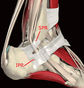
Inferior extensor retinaculum pain or strain can also be accompanied with swelling and tenderness in the ankle region of the foot and the lower legs. Occasionally, the covering that holds the peroneus tendons behind the lateral malleolus (the retinaculum) can be loose or torn and the tendons can snap back and forth out of their normal grooves, this snapping sensation is felt by the patient and can causes further stress/friction on the tendons. Subfibular impingment is seen occurs secondary to calcaneal malunion at the site of torn superior peroneal retinaculum. Also people who stand for prolonged period of time during their job duties are also at risk for injuring or straining the flexor retinaculum of the foot. In a lot of ways, our bodies are strung together like a puppet or marionette, the strings being our tendons, ligaments, and other connective fibers and tissues. Sportsmen involved in running and sprinting can strain or inflame this structure. 22.85±0.94 mm) than cutting of the lateral retinaculum had on medial patellar displacement (lpd: The patella is a sesamoid bone. To view all forums, post or create a new thread, you must be an aapc member. The treatment for superficial digital luxation is a surgical procedure in which the torn retinaculum is sutured back together, thereby restoring the tendon to its correct location. Most of the fibers of the medial patellar retinaculum originate in the medial femoral region from the vastus medialis muscle, just superior to the patella. Tearing of retinacula is more commonly seen at the ankle. Previously torn extensor retinaculum of ankle which is now markedly thickened and irregular (blue arrows).
When the knee moves slightly out of place or becomes tilted in the joint, it can cause tension and pain in the lateral retinaculum. In the painful knee there is a tendency for the patella to tilt toward the outside of the knee. The lateral retinaculum is a ligament that helps hold your patella, or kneecap, in place. If the groove at the point of the heel bone (tuber calcanei) is found to be absent or abnormally shallow, it will be deepened to further increase stability. Likely to be caused by a fatigue tear in the medial capsular insertion into the patella (dutton).
Understanding Subtle Peroneal Subluxation Treating The Cause Vs Symptoms Barefoot Strong Blog from barefootstrongblog.files.wordpress.com The tear is mostly longitudinal split than transverse split. This condition is known as peroneal tendon subluxation or dislocation. The medial patellar retinaculum is the branch of the tendon of insertion of the quadriceps femoris that crosses the knee on the medial side of the patella. Inferior extensor retinaculum pain or strain can also be accompanied with swelling and tenderness in the ankle region of the foot and the lower legs. Does a high grade partial tear or the medial retinaculum in the knee require surgery or is physical therapy a better option? This occurs because of the chronic pull of the knee cap to the outside by the thigh muscles, creating a strain on the medical or inside tissues (the retinaculum). It works with the patella tendon and the quadriceps muscles to help you perform the function of extending your knee. Likely to be caused by a fatigue tear in the medial capsular insertion into the patella (dutton).
Again a previously torn retinaculum will be thickened and ill defined compared to a normal retinaculum which is usually thin and well defined.
They are minor patellar stabilizers and, if intact, can provide knee extension and straight leg raising despite a patellar or quadriceps tendon rupture. When the knee moves slightly out of place or becomes tilted in the joint, it can cause tension and pain in the lateral retinaculum. Also people who stand for prolonged period of time during their job duties are also at risk for injuring or straining the flexor retinaculum of the foot. The patella is a sesamoid bone. Cutting of medial retinaculum had more impact on lateral patellar displacement (lpd: If the groove at the point of the heel bone (tuber calcanei) is found to be absent or abnormally shallow, it will be deepened to further increase stability. The lateral retinaculum is a ligament that helps hold your patella, or kneecap, in place. The flexor retinaculum of the foot can be strained or injured due to a variety of reasons. Subfibular impingment is seen occurs secondary to calcaneal malunion at the site of torn superior peroneal retinaculum. The treatment for superficial digital luxation is a surgical procedure in which the torn retinaculum is sutured back together, thereby restoring the tendon to its correct location. In up to 75% of such patients, lateral ankle. This occurs because of the chronic pull of the knee cap to the outside by the thigh muscles, creating a strain on the medical or inside tissues (the retinaculum). Likely to be caused by a fatigue tear in the medial capsular insertion into the patella (dutton).
Also people who stand for prolonged period of time during their job duties are also at risk for injuring or straining the flexor retinaculum of the foot. The medial retinaculum is located within the knee joint; This condition is known as peroneal tendon subluxation or dislocation. For a torn peroneus brevis tendon, the proximal tenodesis transfers or attaches the brevis to the longus sufficiently proximal to the superior peroneal retinaculum such that the combined tendons at the tenodesis will not pass through the narrow zone of the retinaculum even in maximum inversion. This occurs because of the chronic pull of the knee cap to the outside by the thigh muscles, creating a strain on the medical or inside tissues (the retinaculum).
Cosm Peroneal Tendon Surgery from www.cosm.net.au Inferior extensor retinaculum pain or strain can also be accompanied with swelling and tenderness in the ankle region of the foot and the lower legs. The flexor retinaculum of the foot can be strained or injured due to a variety of reasons. Cutting of medial retinaculum had more impact on lateral patellar displacement (lpd: In up to 75% of such patients, lateral ankle. Subfibular impingment is seen occurs secondary to calcaneal malunion at the site of torn superior peroneal retinaculum. The lateral retinaculum is a ligament that helps hold your patella, or kneecap, in place. The medial and lateral patellar retinaculum are on their respective sides of the patella and are continuous with the vastus fascia to the tibia and the patella. Occasionally, the covering that holds the peroneus tendons behind the lateral malleolus (the retinaculum) can be loose or torn and the tendons can snap back and forth out of their normal grooves, this snapping sensation is felt by the patient and can causes further stress/friction on the tendons.
Stretching this ligament keeps the patella in place and the ligament healthy.stretching a lateral retinaculum of the knee.
Tear of the superior peroneal retinaculum at its attachment to the distal fibula The medial patellar retinaculum is the branch of the tendon of insertion of the quadriceps femoris that crosses the knee on the medial side of the patella. If this is your first visit, be sure to check out the faq & read the forum rules. Again a previously torn retinaculum will be thickened and ill defined compared to a normal retinaculum which is usually thin and well defined. The patella is a sesamoid bone. For a torn peroneus brevis tendon, the proximal tenodesis transfers or attaches the brevis to the longus sufficiently proximal to the superior peroneal retinaculum such that the combined tendons at the tenodesis will not pass through the narrow zone of the retinaculum even in maximum inversion. The medial retinaculum plays a minor role — along with the vastus medialis oblique and the medial patellofemoral ligament — in providing medial stability in. This condition is known as peroneal tendon subluxation or dislocation. Inferior extensor retinaculum pain or strain can also be accompanied with swelling and tenderness in the ankle region of the foot and the lower legs. The treatment for superficial digital luxation is a surgical procedure in which the torn retinaculum is sutured back together, thereby restoring the tendon to its correct location. Most of the fibers of the medial patellar retinaculum originate in the medial femoral region from the vastus medialis muscle, just superior to the patella. In up to 75% of such patients, lateral ankle. If the peroneal retinaculum is torn due to any injury, then the peroneal tendons move out of their place and slip over the lateral malleolus on the external part of the ankle.
If the peroneal retinaculum is torn due to any injury, then the peroneal tendons move out of their place and slip over the lateral malleolus on the external part of the ankle torn retina. They are minor patellar stabilizers and, if intact, can provide knee extension and straight leg raising despite a patellar or quadriceps tendon rupture.


0 Komentar